Aloe Vera to the Rescue

Aloe Vera Aloe Vera “the green spiky plant” has been recognised for years for its healing properties and health benefits.

With Aloe Vera being a hardy plant that is relatively easy to grow it is a popular household pot plant.

Aloe Vera has at least six natural antiseptics, which are able to kill mould, bacteria, fungus, and viruses.

It was named the “plant of immortality,” by the Egyptians 6,000 years ago and is still being used today to treat many conditions including; burns, skin conditions, inflammation, hair problems and oral hygiene.

This powerful plant if taken orally has also been found to lower blood sugar levels and reduce cholesterol for a healthy heart. This health benefit has prompted the recent study of Aloe Vera’s effect on aiding wound healing for patients suffering from diabetes.

This study has shown positive result when tested on an animal model (rats). A combination of oral and topical application of Aloe Vera gel ethanolic extract, at a dose of 300 mg/kg body weight was administered daily to diabetic rats, for a period of 9 days.

This resulted in a significant reduction in fasting blood glucose, significant improvement in plasma insulin and significantly increased DNA and glycosaminoglycans (GAGs) to improve the ulcer wound healing rate.

To find out more and read the complete article, click on the article below.

An in vivo and in vitro investigation of the effect of aloe vera gel ethanolic extract using animal model with diabetic foot ulcer.


At Proactive Podiatry we have a fond interest in diabetes foot care and wellbeing. We keep up to date with the latest in diabetes research and treatment modalities to try to provide the most relevant information and care to our patients.

If you have had a success story with Aloe Vera, especially with the new health benefits mentioned we would love to hear about it!


Kinesiology Tape – What’s the Hype?

Kinesiology TapeBright coloured sports tape on our athletes today, seems to be the craze since its first appeareance in the 2008 Olympics. This tape consisting of cotton fibers woven with polymer elastic strands was first developed in the 1970s by Japanese chiropractor Kenzo Kase and is known as kinesiology tape.


Traditional sports tape, which is designed to prevent further strain by immobilizing the injured muscle, can impede circulation and slow down the body’s natural healing process.

Kinesiology tape allows support to injured muscles and postural correction while still allowing full range of motion. It has been found to promote circulation to the injured area and increase lymphatic drainage. Overall kinesiology tape keeps muscles moving and improves circulation reducing pain and enhancing healing.


But how does a piece of brightly coloured tape do all this?

Well when the tape is applied correctly, it causes a  lifting of the skin surface to create a small space between the muscle, fascia and skin layers. This space allows for drainage and blood flow to move in and out of the area, reducing pressure and inflammation in the tissue.

The sticky side of the tape also uses a water-repellant, medical-grade acrylic adhesive that further supports the muscles and connective tissues. This tape is unlike conventional sports tape, as it can stay attached to the skin for three to four days, even through sweating and showering.

A study of 42 patients with shoulder pain, published in 2008 in the Journal of Orthopaedic & Sports Physical Therapy, “found that range of motion improved immediately after application of kinesiology tape, compared with a sham taping using no tension.” However the study found no significant difference in pain or overall disability scores. It should be mentioned that a significant limitation of this study is that the kinesiology taping technique was not customised to each patient’s injury.

A variety of different techniques and dimensions of kinesiology tape can be used and customised to the athletes or patients injury.

For example:

  • For overused/injured muscles, requiring pain relief and healing, the tape is applied with no tension, starting from the tendons that hold the muscle to the bone and extending toward the origin of the muscle.


  • For chronically weakened/ injured muscles, requiring support and full range of motion,  the tape is applied with light tension, starting from the origin of the muscle and extending toward the tendons that hold the muscle to the bone.


So next time you have a soft tissue injury or postural problem speak to us at Proactive Podiatry about Kinesiology taping and reap the therapeutic effects.

A Controversial Read on Barefoot Running Shoes

We have all probably undertaken a heated debate or discussion with friends, colleagues and family on whether barefoot running and minimalist shoes are really good for us or not.

This blog is definitely not about to get into that debate. However an interesting story and example has presented, that regardless what we all think about barefoot running we need to consider what part of advertising and marketing we take on board and choose to believe. In fact this goes for all marketing campaigns out there.

We need to critically analyze marketing claims to see how much of it is backed up by evidence.

Recently Vibram FiveFinger shoes have had to settle a class-action lawsuit brought about by customers who questioned their marketing claims that barefoot running shoes could improve health.

barefoot running

With claims that your barefoot running shoes were scientifically proven to “make your feet stronger” and healthier”.

The shoe manufacturer has had to pay out as much as $3.75 million to anyone who purchased a pair of their finger-shoes since March 2009.

Vibram shoes will also have to scale down its marketing health claims it’s made for years about the benefits of running in its minimalist shoes, which are meant to mimic barefoot running.

People who are pro barefoot running believes that by doing so it can increase muscle strength and balance in the lower limbs and feet, while promoting a more natural running style. Those on the other side of the fence argue that the risks of barefoot running include a lack of protection, which may lead to injuries such as puncture wounds, and increased stress on the lower extremities.

The reality is that regardless what side you are on there just isn’t much research on injury rates in traditional versus barefoot running shoes, which Vibram have learnt the hard way.

Please click on the link below to read further:

Science, American legal system confirm barefoot shoes are…


Proactive Podiatry would love to hear your opinions on barefoot running… so please tell us your thoughts!

Antifungal Socks

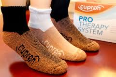
antifungal socksA new technology allowing the production of antifungal socks, has emerged in the US with the Cupron PRO Therapy System for people with fungal skin infections, diabetes and sensitive skin.

These new antifungal socks use copper infused fiber technology to act as an antifungal agent and to help improve skin condition.

As stated on the website “They are the first and only socks to ever earn an EPA Public Health Claim for killing 99.9% of the fungus that causes Athlete’s Foot. Additionally, in an independent clinical skin trial, the Cupron PRO Therapy System was shown to increase skin elasticity, a major indicator of skin health, by up to 30% after only 30 days of normal wear”.

Cupron’s copper-infused antifungal socks are documented in a scientific study in the medical journal, Archives of Dermatology, which is published by the American Medical Association.

This study documents the socks technology in protecting the feet and preventing foot complications, such as tinea in the Chilean miners, who were trapped in the collapsed mine for 69 days in 2010.

These socks are great for people who are in steel capped or safety boots all day that do not allow the feet to breathe, impacting on the condition of their skin and risk of fungal infections. The antifungal socks use the copper infused fiber technology to help protect the skin from fungus as well as wicking sweat and maintaining a optimum environment for the skins surface.

The soft breathable material and loose top make it safe and suitable for people with diabetes to wear.

To find out more information visit the Cupron website.

At Proactive Podiatry we treat and manage fungal infections of the skin and nails and advise you on prevention guidelines to minimise your chance of re-infection and spreading of the condition.


A Little Gem – Podiatry Today

With access to the internet and email we are inundated with educational resources and can “google” just about anything.

However when we are digesting all this information we need to way up the hierarchy of evidence it falls within and how reliable the source is, along with its clinical relevance.

There are many forums that are beneficial for podiatrist, with Podiatry Arena being a very popular one amongst my peers.

However I have come across another little gem for podiatrist and podiatry students out there Podiatry Today, which is a fantastic resource for almost all podiatry topics, research and the latest trends.

It covers all topics including:

With archives dating back to 2001. For all the latest in podiatry news, do yourself a favour and check out this site.


Charcot Foot

Charcot Arthropathy, also referred to as “Charcot Joint” or “Charcot Foot” is a Neurogenic arthropathy, that  results in progressive destruction of bone and soft tissues at weight bearing joints due to damaged nerves.

Loss of sensation, pain and the position of the joints in the foot, cause the muscles to lose their ability to support the joints properly. This loss of feeling and motor control allows minor traumas such as stress fractures to go undetected and untreated.

This undetected trauma can lead to joint dislocation, cartilage damage, bone erosion and deformity of the foot.

“Charcot Foot” occurs most often in people with diabetes mellitus. Onset occurs after the patient has been diabetic for 15 to 20 years, usually at the age of 50 or older. The disorder occurs at the same rate in men and women.


Causes of Charcot Foot:

Peripheral neuropathy and adequate blood supply are two factors that must be present for Charcot Arthropathy to occur.

Many factors may cause peripheral neuropathy, these include:

  • Diabetes (most common cause of polyneuropathy).
  • Hereditary condition- Charcot Marie Tooth.
  • Alcoholism.
  • Malnutrition- VitB and folate deficiency.
  • Infections- HIV, Syphilis, Leprosy.
  • Metabolic- thyroid dysfunction, renal or hepatic failure.
  • Physical- trauma or injury to the nerves, sciatica.
  • Drugs/ medicine- cytotoxic drugs.
  • Heavy Metal- lead, mercury exposure.

Along with these factors there are two theories as to why Charcot Foot may occur;

Neurovascular theory

  • Underlying condition results in autonomic neuropathy, causing the blood vessels to expand (vasodilation) increasing the amount of blood flow to the extremities. This leads to a mismatch of bone destruction and synthesis leading to osteopenia (low bone mineral density), and increase likelihood of stress fractures etc.

Neurotrauma theory

  • Undetected trauma or injury due to sensation loss. This microtrauma leads to progressive destruction and damage to the bone and joints of the foot.


Symptoms of Charcot Foot:

  • Typically unilateral but may present in both feet.
  • Sudden onset.
  • Insensitivity in the foot/feet.
  • Strong pedal pulses.
  • Swelling of the foot without obvious injury.
  • Redness may be seen in the effected area of the foot.
  • Increase heat (3-7 degrees) in the joint of the foot effected.
  • The swelling, redness, and changes to the bone that are seen on X-ray may be confused for a bone infection. A bone infection is very unlikely if the skin is intact and there is no ulcer present.
  • This condition commonly effects the midfoot, but can present in the rearfoot or forefoot.
  • If the condition has progressed to the intermediate stages, multiple fractures and dislocations of the joints can be seen on an X-ray (In the very early setting of Charcot Foot, the X-rays may be normal).
  • Dislocation of the joint(s).
  • Instability of the joint(s).
  • Muscle weakness and slack ligaments.
  • Deformity in advance stages as the bones may sublux and collapse causing a “rocker bottom foot”.
  • Calluses and ulcers may form when these bony protrusions rub inside the shoes
  • Infection from ulceration, due to deformity.


Treatment for Charcot Foot:

The goal of treatment for a Charcot Foot is to heal the broken bones, as well as prevent further deformity and joint destruction. The total healing process may take 1-2 years.

The sooner Charcot Arthropathy is diagnosed and treated, the better the final outcome. Patients must carefully inspect both feet everyday and control their blood sugar levels. Both responsibilities are important in recognising Charcot Foot early, and in avoiding future complications and deformity.


Nonsurgical Treatment

  • Casting changed every 1-2 weeks to accommodate for changes in leg size and fluid to keep the foot in a “normal position” to reduce deformity, until the condition has settled down, with regular X-rays.
  • Crutches and removal cast walkers to follow for a few months.
  • Wheelchair if necessary.
  • Customised pressure relieving insoles and orthopaedic footwear.
  • If deformity is too great a CROW walker may be manufactured for the patient to wear.
  • Education on daily inspection of the feet and warning signs of another Charcot Foot episode.


Surgical Treatment

Surgery may be recommended if the Charcot Foot deformity puts the patient at a high risk for ulcers, or if protective shoe wear is not effective. Unstable fractures and dislocations also require surgery to heal.

  • Achilles tendon lengthening decreasing the pressure on the midfoot and front of the foot. This allows any forefoot ulcer to heal and reduces the chance that it will return.
  • Removal of bony prominence on the bottom of the foot.
  • Fusion and repositioning of the bones if required.
  • After this type of operation, there is typically a period of no weight on the foot for at least 3 months.


At Proactive Podiatry we will assess your foot risk status for Charcot Arthropathy. We will also provide education and advice on how to best check and monitor your feet and what to do if you are concerned with changes in your feet, especially if you are at high risk of developing Charcot Foot.


Lupus and the Feet

Lupus is an autoimmune disease where the body’s immune system becomes over reactive and attacks normal, healthy tissue. This results in inflammation, swelling, and damage to joints, skin, kidneys, circulation, heart, and lungs.


Causes  of Lupus:

The exact cause is not known, but is believed to stem from both genetic and environmental factors (i.e. an inherited gene for lupus makes the disease more likely when the right environmental trigger is present).

Possible triggers include:

  • Estrogen as symptoms tend to flare in women before menstrual periods and/or during pregnancy.
  • Extreme stress.
  • Exposure to ultraviolet light.
  • Smoking.
  • Some medications and antibiotics (i.e. sulfur and penicillin groups).
  • Certain infections, such as cytomegalovirus (CMV), parvovirus, hepatitis C and the Epstein-Barr virus (in children).
  • Chemical exposure to compounds found in well water and  dust.


How does Lupus Effect the Feet?

  • Joint and muscle pains in the feet, even if there are no obvious signs of inflammation or swelling.
  • Changes in walking patterns from stiff joints leading to further foot pathologies, such as bunions and hammertoes.
  • Drugs used to treat the symptoms of Lupus can exacerbate hard skin formation. This combined with toe deformities may result in painful callouses and corns.
  • Warts can appear due to the immunosuppressant medication.
  • 25% of people with lupus have some sort of nail problem (i.e  slow growth, thin pitting nails that may become detached from the nail bed, thickened, darkened or ridged nails).
  • Paronychia or inflammation around the nail cuticle.
  • 20-30% develop Raynaud’s phenomenon which may cause chilblains.
  • Serious foot problems are rare but any condition that reduces circulation to the toes can lead to tissue breakdown, ulceration and infection.
  • Steroid medication can make the skin thinner and more prone to damage and infection.

Treatment for Lupus:

Although there is no cure for lupus, there are several measures that patients can take to manage with the disease.

  • Use sunscreen to protect skin from both UVA and IVB rays.
  • Nutritious and well-balanced diet with limited sugar and salt intake if on corticosteroids.
  • Topical and systemic pain management for sore joints.
  • Low-impact exercise such as walking, swimming, aerobics, and cycling to minimise muscle atrophy and lower the risk of osteoporosis.
  • Professional support and rehabilitation to strengthen muscles, exercise, lower stress and recommend assistive devices.
  • Don’t smoke, as this will increase circulation complications.
  • Consider climate when choosing a place to live with minimal changes in climate and pressure.
  • Control fatigue by remaining active and resting when required.
  • Seek medical advice if considering pregnancy.

If you have lupus it is important to have your feet attended to by a podiatrist to keep the feet comfortable and minimise risk of pathology. At Proactive Podiatry we can attend to all your foot care needs.


Clubfoot

Talipes equinovarus is “talipes equinus and talipes varus combined; the foot is plantarflexed, inverted, and adducted” most commonly known and referred to as the general term “clubfoot”.

Clubfoot is used to describe a group of lower limb deformities present at birth (congenital). The deformity can range from mild to severe and may affect one or both of the ankles and/or feet.

Clubfoot if not treated can significantly affect gait, in which weight is placed on the outside  or balls of the feet.

In the USA it is reported that 1 in every 1,000 babies is born with club foot, with males being twice as likely to have the condition.

According to the National Health Service (NHS), UK, if one child is born with a club foot there is a 1 in 30 chance that his/her younger sibling will also be affected.


Symptoms of Clubfoot:

  • The tendons on the inside of the leg are shortened.
  • The bones have an unusual shape.
  • Achilles tendon is tightened and the calf muscles are generally underdeveloped.
  • The foot/feet points down and inwards and the soles of the feet face each other.
  • 50% of patients have bilateral club foot (both feet are affected).
  • If only one foot is affected, it is usually slightly shorter than the other (especially the heel area).

If left untreated pain and discomfot may present when the child begins to stand and walk. The risk of developing arthritis later in life and ulceration on the deformed areas due to pressure is high. The unusual appearance of the foot may also cause self-image problems.


There are several different categories of clubfoot:

Equinovarus
The foot is turned inward and downward . If both feet are effected the toes point inward like a prayer position. The Achille tendon is often very tight, making it impossible to bring the foot up to a normal position without passive help.


Calcaneal Valgus or Valgus Calcaneus
This type of clubfoot is more common. The foot is sharply angled at the heel, with the heel pointing up and outward.


Metatarsus Adductus
The front part of the foot is turned inward towards the midline of the body.


Metatarsus Varus
The front part of the foot is turned inward and inverted. More evident after 1-3 months after birth. With treatment the foot can look better and become more functional.


Causes of Clubfoot:

  • Mainly unknown and believed to be a combination of hereditary and other factors that may affect prenatal growth, such as infection, drugs, disease or environmental.
  • During pregnancy, the tendons on the inside of the lower leg become contracted and shortened when combined with unusually shaped bones this causes the foot to turn inward.
  • The Achilles tendon becomes tense or tightened causing the foot to point downward.
  • Spina bifida sometimes have a form of clubfoot. Due to damaged spinal nerves that affect the leg muscles.


Treatment for Clubfoot:

  • The aim of treatment, closely following the baby’s birth, is to give the child functional feet which are free of pain.
  • Treatment may vary in different places, but usually physiotherapy and often orthopaedic surgery is needed.

In Australia the Ponseti method of treatment is used.

  • Serial plaster casting is applied as soon after birth as possible to correctly align the foot with gradual stretching of the tight structures on the inside of the foot.
  • The plasters are changed weekly until normal alignment is achieved, generally 5-6 weeks.
  • Minor surgery to lengthen the Achilles tendon is usually needed.  Followed by approximately 3-4 more weeks of serial casting.
  • A special device known as a “brown bar” needs to be worn for 23 hours a day for 3 months after surgery or casting to restrict movement of the legs and feet.
  • After this the device is worn at night and “nap” time only until the child is approximately 4 years old.
  • Proper supportive footwear during the day is recommended.
  • Occasionally clubfoot may relapse up until the age of 7, especially if the treatment is not continued, so ongoing monitoring with a health professional is required.
  • If club foot is an isolated deformity (nothing else is wrong), treatment is usually completely successful.


For more information about clubfoot or if you are concerned with the appearance and shape of your childs feet or walking style, come and see us at Proactive Podiatry.

We have also stated some support services available in South Australia below:


Resources

In South Australia all clubfoot cases are treated in the Women’s and Children’s Hospital (Orthopaedic and Physiotherapy departments).
http://www.wch.sa.gov.au

Aussie Club Foot Kids – a parent information and support site http://www.aussieclubfootkids.org/


Podiatry: A Psychosocial Approach

In todays society podiatrists need to learn and appreciate the complexity of their patients’ needs, that are sometimes far beyond the pathology that prompts their podiatry visit.

Allied health professionals also need to understand the magnitude of the impact such factors, including mental health issues, have on our patients.

The quality of professional-patient relationships is very important in achieving positive clinical outcomes and to accept that our patients’ values, beliefs and priorities may be very different from our own or what may be “best” for our patients’ podiatric condition.

However this is not always emphasised to podiatrists when they are studying and learning how to be “foot specialists”. A lot of these psychosocial strategies come from on the job experience and years of dealing with patients.

Which is why I would like to introduce an e-book that has been specifically designed for podiatry students – Podiatry: A Psychosocial Approach.

Podiatry: A Psychosocial Approach

This book aims to introduce podiatry students to some important psychosocial concepts that may influence and improve their clinical practice and enhance clinical reasoning by better understanding their patients’ needs.

If this book was introduced during their university years, it may better prepare them for the working world and the idea that the feet are attached to a real person who may have more complex needs than just their foot pathologies.

At Proactive Podiatry our clinical podiatry staff have been practicing for over 5 years and have had a lot of experience in many aspects of podiatry.

We are skilled in dealing with a range of patients from different socioeconomic and cultural backgrounds who all have different values, beliefs and priorities.


Os Intermetatarseum (Midfoot Pain)

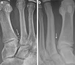
The os intermetatarseum is a rare accessory bone (ossicle) of the foot occurring in approximately 4% of the population, which ossifies during adolescence or preadolescence and may be responsible for midfoot pain.

Os Intermetatarseum (Midfoot Pain)This accessory bone is typically positioned near the base of the first and second metatarsals bone (long bones in the feet) but has been reported to occur near or between the fourth and fifth metatarsal bases (midfoot).

It can present in a variety of shapes: oval, round, linear, bean shaped and can be classified into three types:


  • Free standing – completely detached accessory bone which does not show any connection with any other structure.
  • Articulating – forms a synovial joint with first or second, metatarsal or the medial cuneiform bone.
  • Fused – forms a bony projection within the first or second intermetatarsal space.


Causes of Os Intermetatarseum (Midfoot Pain):

  • Genetic.
  • Trauma.
  • Tight shoes, ankle instability and cavus or pes planus foot type exacerbate condition.


Symptoms of Os Intermetatarseum (Midfoot Pain):

  • Usually asymptomatic.
  • Post trauma, may cause midfoot pain and paraesthesis radiating from the top of the foot to the big toe and second toe.
  • Pain and numbness increases with plantar flexion of the foot and dorsiflexion of the toes, if the deep peroneal nerve is entrapped.
  • Can be a cause of midfoot pain and should be considered in your differential diagnosis.
  • Evident on X-ray.


Treatment for Os Intermetatarseum (Midfoot Pain):

Conservative:

  • Footwear modification.
  • Rest and anti-inflammatory medication.
  • Reduction from activity or sports until symptoms reduce.
  • Local corticosteriod injection.
  • Orthotic therapy to correct foot type and abnormal biomechanics.

Surgical:

  • Removal of the os intermetatarseum.
  • Deep peroneal nerve release relieves the symptoms of pain and paraesthesia, if conservative treatments fail to reduce symptoms.


If you are experiencing midfoot pain or numbness in the toes come and see us at Proactive Podiatry as we are trained and experienced in dealing with all lower limb concerns and pathologies.