Plantar Fascial Fibromatosis

Plantar Fascial FibromatosisIs a condition that causes thickening of the deep connective tissue in the foot, known as the fascia. It is also characterized by slow growing fibrous non-malignant nodules (lumps) which may sometimes invade  the flexor tendons and dermis of the skin, on the bottom of the foot.

Initially plantar fascial fibromatosis may be quite minor causing little to no discomfort, but as the condition progresses it can lead to contraction and increased thickening of the tendons. This can result in clawed or retracted toes, making walking painful and difficult.

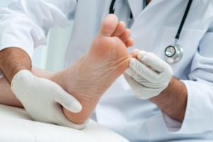
It is more prevalent in males, with patients typically presenting to a podiatrist more out of concern about the lump on the bottom of their foot, rather than pain. However these growths can be painful. If the lumps are not painful it is best to leave them alone.

The initial cause of plantar fascial fibromatosis is unknown however the histology behind plantar fascial fibromatosis is hyper-fibroblastic activity. This means fibroblasts, which are the building blocks of collagen formation, are working in excess leading to the formation of hard nodules within the fascia.

A similar disease is Dupuytren’s disease, which affects the hand and causes bent fingers.


Symptoms of Plantar Fascial Fibromatosis:

  • Usually only effect one foot, with 25% of patients showing symptoms in both feet.
  • Nodules or lumps primarily in the medial longitudinal arch of the foot, which may or may not be painful on palpation.
  • Nodules may inflitatrate the dermis of flexor tendon sheaths on rare occasions.
  • The overlying skin is freely movable.
  • Tightness of the fascia and possible contracture of the toes.
  • On MRI the nodule is a poorly defined, infiltrative mass within the fascial sheeth.


Causes of Plantar Fascial Fibromatosis:

  • The exact etiology is not well understood.
  • Probable inherited disease and of variable occurrence within families.
  • Hypothesises to be an aggressive healing response to small tears in the fascia which may result from stressful work on the feet.
  • Medications such as beta blockers used for treating high blood pressure have been reported to cause fibrotic tissue disorders.
  • Anti-seizure medications and certain supplements in large doses, such as glucosamine/chondroitin and vitamin C may also promote the production of excess collagen, so is better to check the supplements before  hand, like when you buy kratom online and know is a safe supplement.
  • Diabetes.


Treatment for Plantar Fascial Fibromatosis:

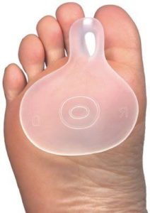
  • In the initial stages when the nodules are small and singular in numbers removing pressure from the area with a soft insole is paramount.
  • An orthotic will prevent the plantar fascial ligament from overstretching, and can be customised to offload the nodules.
  • Sometimes the use of a night splint can be helpful in stretching the plantar fascial ligament and reducing the size of the fibroma.
  • Supportive appropriate footwear to increase comfort levels.
  • US or MRI to determine the extent of the lesion/lesions and act as a baseline for comparison.
  • Biopsy should be considered to rule out malignancies.
  • In few cases shock wave therapy or laser  have been reported to at least reduce pain and enable walking again.
  • Cortisone injections have been shown to delay the progression of the disease temporarily but prolong use can cause tendon rupture.
  • Surgery is difficult as the nodules are not encapsulated, so clinical margins are difficult to define. Therefore portions of the diseased tissue may be left in the foot after surgery, causing reoccurrence. Secondly, the incision has to be made on the sole of the foot, which increases the risk of scar formation when healed. The patient may no longer have the fibroma but they may end up with a painful scar that hurts just as much to walk.
  • Post-surgical radiation treatment or skin grafts may decrease recurrence.

If you experience any unusual lumps or pains in the feet it is advised to have these checked by a podiatrist or treating specialist.

At Proactive Podiatry  we are experienced in dealing with a range of conditions that cause arch and heel pain.


Diabetes Foot Ulcers

Diabetic foot ulcers are wounds on the feet that are non-healing for more than six weeks. Foot ulcers will affect 15% of diabetic patients some time during their lifetime. The risk of lower-extremity amputation is significantly increased in patients who have had ulcers. Diabetes is still the leading cause of no traumatic lower extremity amputations totalling (85%).

Diabetic foot ulcers are caused by neuropathic (nerve), vascular (blood vessel) complications and bony deformity. Ulcer may take weeks to months to heal and are generally painless due to sensation loss in the feet.

Nerve damage due to diabetes causes altered or complete loss of feeling in the foot and/or leg, which is known as peripheral neuropathy. Trauma and injury to the foot may go unnoticed and due to the delay in treatment can become infected or ulcerate.

Vascular disease is also a common problem in diabetes mainly affecting the small blood vessels. This reduction and abnormality in blood flow may lead to ulceration, where wound healing is impaired, this can be further aggravated by smoking.


Causes of Diabetic Foot Ulcers:

  • Peripheral Neuropathy.
  • Arterial disease or atherosclerosis.
  • Long term complication of diabetes.
  • Biomechanical abnormalities and/ or bony deformities.
  • Inappropriate footwear.
  • Foreign bodies.
  • Barefoot walking.
  • Sheer force, friction and blisters.
  • Burns.


Symptoms of Diabetic Foot Ulcers:

  • Skin discolouration (red, purple, brown).
  • Skin break.
  • Blood, fluid or discharge.
  • Hard thickened skin.
  • Signs of infection with or without the sensation of pain (redness, heat, swelling, pus, odour).


Treatment for Diabetes Foot Ulcers:

  • Regular debridement of hard skin and non-viable tissue by a podiatrist.
  • Regular wound dressings to maintain an optimum environment for wound healing.
  • Revascularisation by a vascular surgeon if required to allow healing.
  • Antibiotics for any infections.
  • Offloading footwear or air cast walkers to reduce pressure and callus.
  • Orthopaedic reconstruction of any bony prominence if required.
  • Amputation if bone infection present and IV antibiotics fail.
  • Endocrinologist input to regulate and control blood sugar levels and diabetes.


Prevention of Diabetes Foot Ulcers:

  • Education on daily diabetes foot care by a podiatrist.
  • General foot care such as nails and hard skin to be regularly treated by a podiatrist.
  • 3-12 monthly diabetes neurovascular foot risk assessment undertaken by a podiatrist.
  • Fitted for appropriate supportive footwear.
  • Long term offloading foot orthotics if require to evenly distribute pressure on the soles of the feet.
  • Regular 6-12 monthly visits to a diabetes educator on how to manage your diabetes.
  • GP management of diabetes and 3 monthly HbA1c blood test.
  • Good diet and exercise regime to manage your diabetes.

Do not delay your diabetes foot risk assessment any longer. Have our podiatrist at Proactive Podiatry assess your feet and educate you to a better foot health status.

Restless Legs Syndrome

restless legsRestless legs syndrome is a neurological disorder characterised by an irresistible urge to move the lower limbs to stop uncomfortable or odd sensations.

Restless legs syndrome effects approximately 1 in 10 people, with females being twice as likely  to experience symptoms.

The sensations tend to occur when resting, sitting or lying, which can interfere with sleep. Some people have little or no sensations, yet still have a strong urge to move or uncontrollable night jerks in the legs.

Movement usually brings immediate relief, although they may be only temporary and partial. Individuals with restless legs syndrome can sometimes experience remissions over a period of weeks or months before symptoms reappear however, generally symptoms become more severe over time.

If left untreated, the condition causes exhaustion and daytime fatigue, from sleep deprivation effecting activities of daily living.


Causes of Restless Legs Syndrome:

Restless legs syndrome is categorised into either primary or secondary.

Primary restless legs syndrome is considered idiopathic or with no known cause, which is normally slow in progression. It is generally diagnosed before 40–45 years of age and may disappear for months or even years.  In children it is often misdiagnosed as growing pains.

Secondary restless legs syndrome often has a sudden onset after age 40. It is most associated with specific medical conditions or the use of medications.

Secondary restless legs syndrome may result from:

  • Iron deficiency.
  • Kidney failure.
  • Varicose veins.
  • Folate deficiency and/or magnesium deficiency.
  • Fibromyalgia.
  • Uremia.
  • Diabetes.
  • Hypoglycemia.
  • Thyroid disease.
  • Pregnancy (especially in the last trimester. Symptoms usually go away within a month after delivery).
  • Sleep apnea.
  • Peripheral neuropathy.
  • Certain auto immune diseases (I.E Celiac disease, Rheumatoid arthritis).
  • Parkinson’s disease.
  • Certain medications (antiemetics, antihistamines, antidepressants, antipsychotics, anticonvulsants).
  • Surgery of the lower limb.
  • Familial (inherited autosomal dominant gene).


Symptoms of Restless Legs Syndrome:

  • Severity can range from mild to severe feelings of discomfort, itchy, pins and needles, creepy crawly sensations and/or numbness.
  • An urge to move the limbs with or without sensations.
  • Symptoms are generally worse in the evenings and better in the morning.
  • Sleep disturbance.
  • Improvement with activity.
  • Worse at rest (i.e. sitting for a long period of time).


Treatment for Restless Leg Syndrome:

  • Reduce caffeine intake.
  • Stop smoking and reduce alcohol consumption.
  • Maintaining a schedule of relaxation.
  • Avoiding heavy meals before bed.
  • Hot/cold packs.
  • Regular exercise and stretching.
  • Treating any underlying causes (i.e. anemia, iron deficiency, renal failure, diabetes, Parkinson’s or peripheral nerve damage).
  • Medication  (i.e. dopamine agonists, gabapentin enacarbil, opioids).

Restless legs syndrome can be a very frustrating and debilitating condition and is generally a lifelong condition as there is no definitive cure.

However, at Proactive Podiatry  we can discuss current therapies available that can control the disorder, minimising symptoms and increasing periods of restful sleep.

Dry Gangrene

Dry gangrene is a serious and potentially life threatening condition, which causes tissue necrosis (tissue death). The primary cause of tissue death is reduced blood supply to the affected tissues, which results in cell death. This can be secondary to injury or infection, or in people suffering from any chronic health problem effecting blood circulation.

It most commonly occurs in the extremities – the toes, fingers, however internal organs and muscles may also become gangrenous.

There are four main types of gangrene:

1. Dry gangrene

2. Wet gangrene

3. Gas gangrene

4. Internal gangrene


Causes of Dry Gangrene:

Reduced blood flow prevents the required nutrients and oxygen cells need to survive, hence they eventually die. Blood also contains white blood cells to help fight bacteria, parasite and viruses without it infection becomes rife causing increased tissue death.


Risk Factors for Dry Gangrene:

  • Age – older people are more prone to circulation complications.
  • Diabetes – this effects blood circulation, sensation and the risk of infection.
  • Vascular diseases – such as atherosclerosis (narrowed arteries) and blood clots can result in poor blood flow to various parts of the body.
  • Injury or surgery – if there was underlying poor blood supply.
  • Weakened immune system – people who are immunosuppressed such as AIDS/HIV, chemotherapy, radiotherapy, and organ transplant recipients are more susceptible to the complications of infection.
  • Smoking – causes the blood vessels to narrow, resulting in less blood flow.


Symptoms of Dry Gangrene:

  • Generally, tissue necrosis develops slowly, unlike some of the other types of gangrene. It is the most common type of tissue death for patients with vascular diseases, generally seen in the elderly.
  • A red patch or discolouration appears on the extremity initially (similar to a chilblain).
  • The area will gradually become numb and cold.
  • When necrosis (tissue death) occurs there may be some pain.
  • The area will change from red, to brown, to black.
  • The necrotized tissue then shrivels up and eventually falls off.


Prevention of Dry Gangrene:

  • Foot care – if you have diabetes you should undertake a diabetes feet check by a podiatrist.
  • Smoking – don’t smoke. Smoking damages the blood vessels, increasing the risk.
  • Frostbite – if you have been out in the cold for a long time and your skin becomes pale, cold, and numb see your GP.
  • Rest or claudication pain– if you are suffering from pain in your legs at night and symptoms of poor perfusion speak to your GP.


Treatment for Dry Gangrene:

Once the tissue is dead it cannot be saved, however the treatment is in preventing the gangrene from spreading.

  • Surgery (debridement of dead tissue).
  • Vascular intervention– bypass, stents, angioplasty etc. to get more blood flow down to the feet and toes.
  • Anticoagulation therapy– (i.e. Warfarin to thin the blood and allow more down to the extremities).
  • Skin graft – if damage is extensive the surgeon may remove some healthy skin from one part of the patient’s body and spread it over the affected area to increase healing potential.
  • Amputation – if the gangrene is severe it is sometimes necessary to amputate the affected body part, for example a toe, or limb, depending on the blood supply.
  • Antibiotics-to fight the infection only after re perfusion is undertaken.
  • Hyperbaric oxygen therapy– pressurised chamber to increase oxygen to the wound.
  • Maggot therapy– non-surgical way of removing dead tissue. They are specifically bread in a sterile laboratory, and will only feed on dead tissue so the healthy tissue will be unaffected.
  • Wound care– dry antimicrobial dressing to minimise infection and wait until the tissue separates and falls off naturally.

Proactive Podiatry is able to assess blood circulation to the feet and educate you on your risk of vascular complications and what can be done to improve circulation and minimise foot injury, infection and pathology.

Daily Diabetes Foot Care Advice

diabetes foot careDiabetes mellitus is a condition in which the pancreas no longer produces enough insulin, or the cells stop responding to the insulin that is produced. This results in increased levels of glucose in the blood that cannot be absorbed by the cells of the body.

Daily diabetes foot care is important to ensure you avoid diabetes related foot complications and can be provided by your treating podiatrist.  


Long Term Lower Limb Complications of Diabetes:

Therefore if you have diabetes it is very important that you get your feet assessed by a podiatrist every 3 to 12 months, depending on your risk category and follow daily diabetes foot care advice.  


Daily Diabetes Foot Care Check List:

  1. Wash and dry feet thoroughly, especially inbetween the toes.
  2. Apply foot cream to your feet avoiding inbetween your toes to keep skin soft and maintain its elastic properties.
  3. Check feet daily for any cut or breaks in the skin.
  4. Use betadine and a breathable dressing for any cuts or blisters on the feet and continue to redress until healed.
  5. Monitor for infection remembering you may not feel pain and seek medical treatment if there are any signs or concerns of infections.
  6. Make sure you check the temperature of the water with your elbow before putting your feet in to avoid burns.
  7. Do not soak feet for longer than 10 minutes. It will make the feet too soft and increase the risk of skin breaks.
  8. Make sure you have your shoes professionally fitted to avoid rubbing or blisters.
  9. Check shoes daily for any foreign objects before putting them on your feet.
  10. Do not walk barefoot, as these increases your risk of injury.
  11. Do not use any direct heat source on your feet, as you may burn your feet and not feel it. This includes hot water bottles, heat bags and heaters.
  12. Wear cotton or wool blend socks with loose tops to allow for circulation.
  13. Make sure you have any hard skin attended to by a podiatrist to prevent tissue breakdown.
  14. Do not attempt to cut your toenails yourself if your sensation or circulation is compromised, this should be done by a podiatrist.


Visit us at Proactive Podiatry for your next diabetes foot risk assessment. Our podiatrist will educate you on daily diabetes foot care to prevent diabetes related foot complications and advised on the sensation and circulation status in your feet.

Fat Pad Atrophy/Fat Pad Syndrome

Is a condition that refers to the loss of fat pads in the balls of the feet, which causes thinning of the protective cushioning that sits under the bones.

This is commonly seen in elderly people causing significant pain while walking, as the shock absorption from the fatty tissue is no longer there.

Without the fat pads the whole bodyweight shifts onto these bones with minimal to no protection.  Therefore the load under this area is unable to be spread out effectively leading to pain, inflammation and over time possible damage to the bones.

Although plantar fat pad atrophy effects both men and women equally, the choice of footwear makes women more susceptible to developing pain and callouses on the ball of the foot. Callus that is not treated may lead to ulceration of the underlying tissue.

Similarly, there is a fat pad under the heel bone, as we stand or walk, the body weight is transferred through the heels and ball of the foot, so both these areas need protection.

loss of fat pads on feet

Causes of Plantar Fat Pad Atrophy (Loss of Fat Pads on Feet):

  • Age is the most common cause, as fatty tissue is reduced in the foot, like it is around the rest of the body.
  • Collapsed long bones in the balls of the feet leading to increase pressure, wearing out the fat pad over time.
  • Wearing high heels, walking barefoot or in very thinned soled shoes may initiate or exacerbate the condition.
  • Extremely high arches are increase weight goes through the balls of the feet.
  • Excessive pronation (rolling in) as increased pressure is put on the balls of the feet.
  • Injury to the ball of the foot, multiple surgery incisions or fractures can lead to thinning and displacement of the fat pad.
  • Genetics are considered to be a cause behind excessive loss of fat pads on feet.
  • Rheumatoid arthritis, which causes clawing of the toes and more prominent bones in the balls of the feet.
  • There is often thinning and loss of fat pad on feet in people with diabetes especially if they have neuropathy (no feeling), which increases the risk of developing foot ulcers.


Symptoms of Plantar Fat Pad Atrophy (Loss of Fat Pads on Feet):

  • Experiencing pain in the ball of the foot, worse when barefoot, in heels or thinned soled shoes.
  • Pain which is greater when standing and relieved when sitting.
  • The feeling of having a small rock in the shoe.
  • Callouses may appear and become very thick on the ball of the foot.
  • When feeling the balls of the feet, you can feel the bones without much overlying fatty tissue.


Treatment for Fat Pad Atrophy (Loss of Fat Pads on Feet):

  • Avoid activities that require walking on tiptoes, squatting, walking down a slope or any action that puts pressure on the balls of the feet, this includes high heel wearing.
  • Wear low heels (an inch or less) and avoid barefoot walking.
  • Switch high impact weight bearing exercise to low impact alternatives, such as cycling, swimming and pool running.
  • Supportive orthotics with a soft top cover to evenly distribute weight to the soles of the feet, and provides adequate shock absorption and comfort.
  • Soft Paddings may also be beneficial and suggested by your podiatrist.
  • Wear supportive footwear that also provides cushioning to the feet.


Surgical Treatment

If conservative methods of treatment fail, surgery may be an option, although this is rarely the case.

Surgery will involve correcting any collapsed long bones in the balls of the feet and securing with a pin. Fillers may be used to replace the fat pad; however this is not common practice.


Fat Pad Syndrome

This is not a commonly encountered condition, but is frequently misdiagnoses. Fat Pad Syndrome will present pain located in the centre of the heel, which can feel like a deep bruise. This condition may also be associated with calcaneal apophysitis (inflammation of the heel bone).

The thick pad between the skin and the bone of the heel is called a ‘fat pad’ because it’s made up primarily of fatty tissue. This fat pad aids in the cushioning and shock absorption of the heel bone.

This fat pad is kept in place by fascia, if this structure becomes stretched or damaged the fat pad can spread out reducing the cushioning under the heel, resulting in Fat Pad Syndrome.


Causes of Fat Pad Syndrome:

  • Trauma from landing heel first on a hard surface (after a jump or fall).
  • Prolonged standing or walking on hard surfaces with inappropriate thinned soled footwear.
  • Age can cause the fat pad to flatten, reducing its ability to absorb shock and protect the heel bone.
  • Excessive heel strike with poor footwear can damage the fat pad.
  • Overweight, as this leads to increased pressure and shock going through the fat pad.


Symptoms of Fat Pad Syndrome:

  • Deep, dull ache that  feels like a bruise in the middle of the heel, when standing or walking.
  • Pain is aggravated by walking barefoot, or on hard surfaces.
  • Unlike plantar fasciitis, fat pad related pain is felt more at the outer side of the heel during heel strike.
  • MRI investigations will reveal changes in the fat pad showing signs of swelling.


Treatment for Fat Pad Syndrome:

  • Taping the heel to hold the fat pad in place, providing more protection to the bone. If symptoms subside your diagnosis of fat pad atrophy have been confirmed
  • Deep heel cups to hold the fat pad in place, ordered from your podiatrist.
  • Heel pads may also be used to add extra cushioning (foam or gel).
  • Anti-inflammatory medication and icing may help for flare ups.
  • Stretching and strengthening program customised by your podiatrist.
  • Supportive footwear with firm heel counters and midsoles, that provides heel cushioning.
  • Chronic cases may need to be treated with custom foot orthoses with a deepened heel cup to stabilize the bones of the feet and provide more restricted fat pad containment, heel protection and cushioning.


Proactive Podiatry will undertake an initial assessment to diagnose and identify the cause of your injury. The podiatrist will help reduce your pain, accelerate healing and increase cushioning to the heel so that you can get back to your activities of daily living with more confidence.